

Advanced Corneal Imaging and Keratoconus Monitoring
Modern keratoconus management relies heavily on advanced corneal imaging technologies.
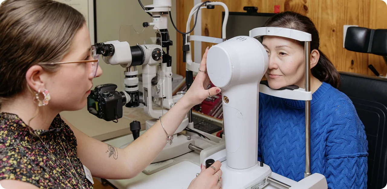
At Rose Optometry and the New Zealand Eye Research Centre (NZERC), multiple advanced imaging systems are used to monitor corneal shape, detect progression and guide treatment decisions.
Tomography is considered the gold standard for keratoconus monitoring and severity assessment because it analyses both the front and back surfaces of the cornea together with corneal thickness distribution.
Technologies used include:
Modern tomography systems allow clinicians to identify subtle posterior corneal changes before advanced visual symptoms develop.
This enables earlier intervention including:
- Corneal cross-linking
- Specialty contact lenses
- Long-term progression monitoring
Hamilton’s Role in Global Keratoconus Care
Hamilton, New Zealand has played an internationally significant role in the history of keratoconus management.
Rose Optometry at 38 Lake Road was founded by Paul Rose, developer of the internationally recognised Rose K keratoconus lens design.
The Rose K system became one of the most widely used specialty keratoconus lens systems globally and contributed significantly to modern rigid lens fitting philosophy.
Today, Rose Optometry and NZERC continue this legacy through:
- Advanced corneal imaging
- Scleral lens fitting
- Orthokeratology
- Keratoconus research
- Ectasia management
- Screening advocacy
- Specialty contact lens innovation
The clinic accepts national and international referrals for complex corneal and contact lens management.
National Keratoconus Screening Advocacy
New Zealand has one of the highest documented rates of keratoconus internationally, particularly within Māori and Pacific communities.
Research from New Zealand has demonstrated significantly elevated prevalence rates in adolescents, supporting the need for earlier detection strategies.
Rose Optometry and NZERC advocate for improved keratoconus screening because:
- Keratoconus commonly begins during adolescence
- Progression is fastest in teenage years
- Early treatment improves outcomes
- Corneal cross-linking is most effective before advanced progression occurs
Modern corneal tomography systems now allow detection of subtle ectatic changes before severe vision loss develops.
The goals of future screening programmes include:
- Reducing preventable visual impairment
- Decreasing corneal transplantation
- Improving equity of care
- Identifying disease earlier
- Improving long-term visual outcomes
References
- Papali i-Curtin AT et al. Keratoconus prevalence among high school students in New Zealand. ʻ Cornea. 2019.
- Gokul A et al. Aotearoa Research into Keratoconus (ARK) Study. Cornea. 2022.
- Gomes JAP et al. Global Consensus on Keratoconus and Ectatic Diseases. Cornea. 2015.
- Hong CY et al. Screening plus corneal cross-linking for keratoconus is cost-effective for New Zealand. Clinical and Experimental Ophthalmology. 2025.


.svg)

